Heart MRI
Euromedica General Clinic of Thessaloniki in cooperation with EVZOIA have developed and now operate, the specialized facilities for conducting magnetic resonance imaging of the heart (or Heart MRI). An extremely important and reliable imaging method to assist in the diagnosis and treatment of heart conditions, the provision of which until now has been particularly limited in the city of Thessaloniki.
What is a heart MRI?
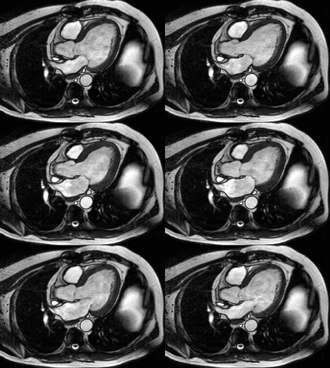
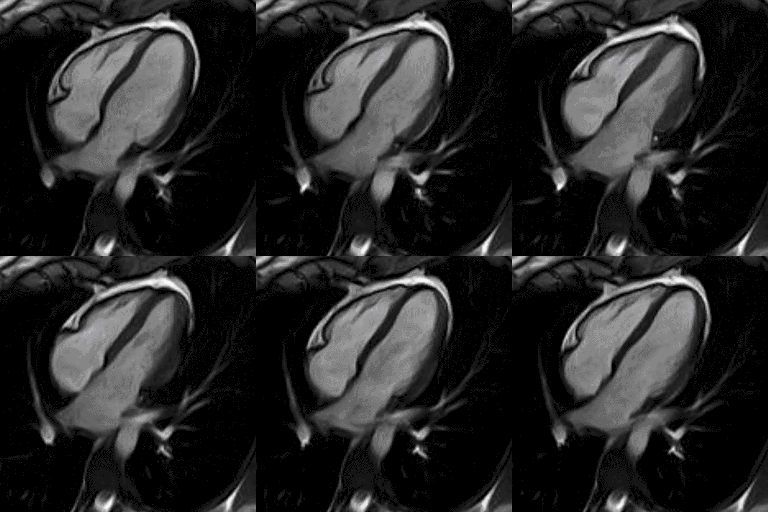
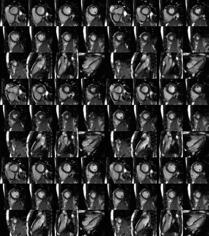
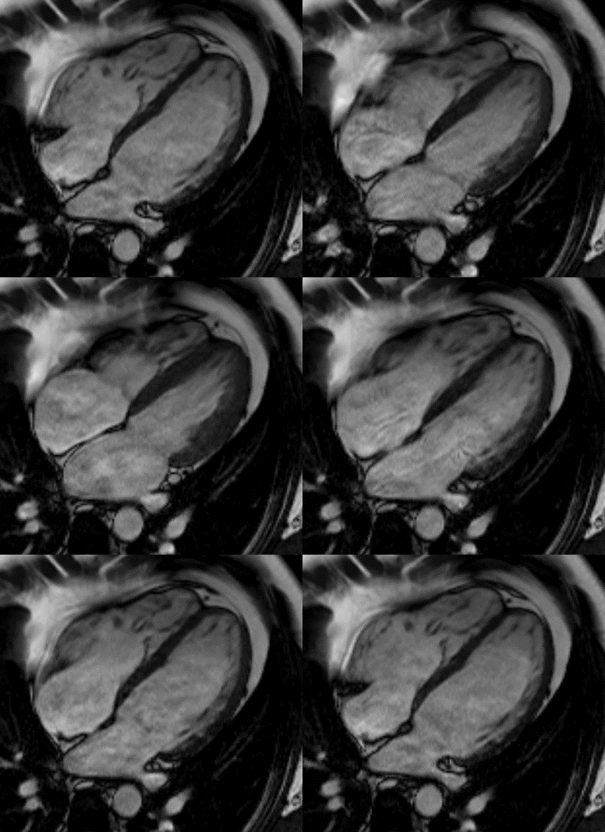
 A heart MRI is a specialized medical imaging method for a non-invasive assessment of the morphology and heart function. It uses magnetic waves under electrocardiographic resonance, to produce high-resolution static and dynamic images of the heart and its large vessels, without the use of ionizing radiation. Based on these images, diagnosis can be performed with high reliability and lead to a detailed treatment for most heart conditions.
In which cases is a Heart MRI indicated?
 A heart MRI is considered today the only non-invasive method for diagnosing myocarditis, both acute and chronic. Apart from myocarditis, it is indicated and contributes significantly to the investigation and diagnosis of cases such as:
- ischemic cardiomyopathy
- invasive cardiomyopathies
- congenital but also acquired, non-ischemic cardiac myocardiopathies
- heart failure
- arterial and pulmonary hypertension
- pericardial diseases
- cardiac valvular diseases
- myocardial iron load on hemoglobinopathies
- cardiac masses
- congenital heart abnormalities
- coronary artery abnormalities
- diseases of the large vessels
- postoperative results after cardiac surgery
A heart MRI is also indicated to those who do not have a known cardiological problem, but maintain some concerns about the good health of their heart. And as it is the most important organ of the human body, the non-use of ionizing radiation is a given positive.
How is a heart MRI conducted
 The test takes about 40-45 minutes. During the examination, the patient is lying down, facing upwards on the magnetic tomography table, listening to music of their choice. Meanwhile there is constant visual and acoustic communication with the MRI scanner technitian, for anything the patitent may need.
Sometimes the patient will be asked to hold their breath for a few seconds following clear instructions from the technitian, to maximize the quality of the heart images produced. The patient’s arm is fitted with a venous catheter for the delivery of the contrast medium, which is absolutely essential for the more detailed imaging of the anatomy, but also for possible pathological findings of the heart.
Advantages of a Heart MRI
 A Heart MRI today, has many and undoubtedly important advantages that make it a particularly powerful diagnostic and prognostic tool, in the field of modern cardiology. Some of them are the following:
- Non invasive
- Absolutely safe and harmless to the human body, given its non-ionizing radiation
- High-resolution imaging of the heart’s anatomy and its large vessels, superior to the other available imaging methods High-resolution imaging of the anatomy of the heart and its large vessels superior to the other available imaging methods
- Detailed study of morphology and heart function, superior to the other methods available
- Ability to investigate and diagnose the majority of heart diseases, congenial or aquired
Supplemental and particularly valuable, in cases where other diagnostic methods are appropriate
Ideal even in the case of prophylactic testing for heart diseases
Financial coverage of the examination by the national insurer
Limitations of a heart MRI
The same ones that apply to any magnetic resonance imaging examination.
